Pathogens Detection via MALDI-TOF MS Technology

One of the most commonly used methods for microbial identification is 18S and 16S ribosomal RNA sequencing. Currently a new technology, MALDI-TOF MS (matrix-assisted laser desorption ionization-time of flight mass spectrophotometry) has emerged as a potential technique for the diagnosis of microbes in clinical specimens. This technique is very rapid, has high specificity and sensitivity as compared to other conventional methods of pathogen identification. Microbiologists have used this technique not only for diagnostic purposes but also in epidemiological studies, strain identification and typing, detection of toxins, identification of biological warfare agents, water, and foodborne pathogen, detection of antimicrobial resistance, and identification of urinary tract and blood pathogen.

Working Principle

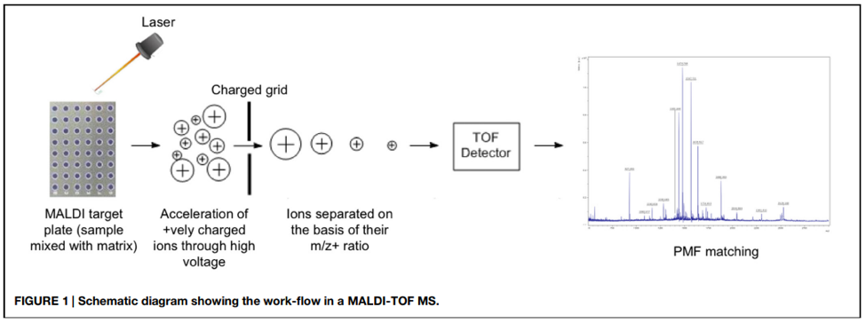
The specimen is mixed with a matrix substance and placed onto the surface of the MALDI target plate. The matrix is allowed to dry to crystallize the sample along with it. The plate is then loaded into the machine where each sample is bombarded with short laser beams. The sample becomes vaporized due to laser energy which generates ionized protein (ribosomal) particles. An electromagnetic field accelerates these particles prior to their entrance into the vacuum-generated flight tube. The time taken by each ionized particle to reach the end of the flight tube is measured precisely which produces a unique peptide mass fingerprint (PMF) for each analyte in the specimen. The PMF is compared with information available in the databases to make a conclusion about pathogen detection (Figure 1).

Almost all types of microbial pathogens in stool samples, blood cultures, urinary tract infections, respiratory tract infections, and cerebrospinal fluids can be detected with MALDI-TOF MS.

The sample preparation before its analysis via MALDI-TOF depends upon its chemical composition and sources from which it was obtained.  Different sample preparation methods have been evaluated by the scientists. Some pathogens can be identified directly from the specimen, also called direct cell profiling, while some pathogens need to be purified and their crude cell extracts are prepared.

Detection of bacterial and fungal pathogens grown on agar plates with MALDI-TOF MS

The conventional methods of bacterial identification are time-taking and laborious. MALDI-TOF MS analysis can reduce the time up to 30 minutes if samples are already prepared, specifically if host contamination is removed from the sample. Almost all types of pathogenic bacteria both gram-positive and gram-negative e.g., Listeria monocytogenes, Burkholderia mallei, Enterococcus faecalis, Clostridium botulinum, Corynebacterium diphtheriae, Bacillus cereus, Brucella melitensis, Corynebacterium ulcerans, Francisella tularaensis, Shigella dysenteriae, Vibrio cholerae, Yersinia pestis, Streptococcus pyogenes, and Legionella pneumophila can be detected by MALDI-TOF MS.

The specimens for bacterial detection are first grown on an appropriate selective media. The colonies are subjected to protein extraction via either chloroform extraction, trifluoroacetic acid extraction, or acetonitrile extraction method. The extracted proteins are analyzed with MALDI-TOF MS for bacterial identification. The bacterial culture should be as fresh as possible because ribosomal protein degradation occurs in older cultures which may lead to inefficient species identification.

Direct analysis of blood for the identification of bacteria with MALDI-TOF MS

Blood pathogens can be detected with MALDI-TOF MS faster if first remove the host body proteins which interfere with the detection of pathogens. Removal of host body proetins is crutial as it removes the background noise and makes the MALDI TOF MS analysis more efficient, otherwise it may end up with false positive results. The Devin Fractionation filter is a good solution in this way as it is able to remove most of host contamination (> 95%) in just 5 minutes and has a very high microbial passing efficiency (>99%). So it can be easily integrated in most routinely used methods of sample preparation in all of in-house methods.

Urine analysis for bacterial pathogen detection with MALDI-TOF MS

Urine analysis for the detection of pathogens can be performed by MALDI-TOF MS. It is quite challenging to analyze a sample directly because samples contain host normal flora and proteins which can hinder the mass spectrum of the pathogen. However, urine is a unique sample amongst all as it does not contain host proteins and normal microflora. Further, urinary tract infection is often monomicrobial and represents a higher concentration of the microbes during the infection.  A fast centrifugation step will pallet down all the bacteria in the sample, which is washed with distilled water to remove any residues. The pallet of the bacteria is applied onto the surface of the metallic MALDI plate. 91.8% species-level identification can be carried out with urine samples.

MALDI-TOF MS in Clinical Virology

MALDI-TOF MS technique has also been used by microbiologists to detect viral pathogens in clinical specimens. The gold standard method for viral detection is cell culturing which requires a very sophisticated setup and takes a lot of time. The applications of MALDI-TOF MS technology in clinical virology are less as compared to mycology and bacteriology, which is because of the fact that viruses have low protein content. However, the applied aspect of MALDI-TOD MS has been proved by virologists. The human enteroviruses, influenza virus, herpes virus, human papillomavirus, and hepatitis virus have been detected via this technique. For this, viral genetic material should be first extracted; for efficient DNA extraction Devinâ Microbial DNA enrichment kit offers optimized sample preparation and enrichment within 1.5 hours! After amplification, the amplicons are analyzed with MALDI-TOF MS. Recently, modifications have made it possible to detect multiple viral pathogens simultaneously from the specimens with the help of the multiplex MALDI screening method.

Limitations and challenges of MALDI-TOF MS

Efficient clinical diagnosis required fast pathogen detection. With the help of MALDI-TOF MS pathogen can be identified up to species level within 30 minutes from the colony and the results can be consulted with the physician to propose appropriate treatment.

The invention of MALDI-TOF MS has made it possible to replace the conventional methods of pathogen detection and disease diagnosis. However minor discrepancies have been observed in the results of molecular, biochemical, serological, and MALDI-TOF oriented results. The MALDI analysis is based upon the spectrum of ribosomal proteins, so the microbes which do not differ much in their ribosomal protein structure and sequences like E. coli, S. pneumoniae, and Shigella species, are difficult to distinguish. That is why the need for molecular methods and biochemical methods remains for pathogen detection. The high through put technique next generation sequencing (NGS), aligned with bioinformatics is an advanced and powerful technique for pathogen detection and identification. On the other hand, MALDI-TOF MS is time-saving and does not depend on metabolic reactions. Further, contrary to conventional methods, all types of gram-positive and gram-negative bacterial can be detected with a single MALDI-TOF MS system and no prior pre-differentiation is required for species-level identification.

An important challenge for this technique is the development of PMF databased, with which the sample’s data is compared and analyzed. The detection of new strains and isolated is only possible when the PMF of the new strain is already present in the database. It is crucial to improve the quality of the database and introduce quality controls for more accurate pathogen detection. Appropriate calibration and maintenance of the device are also required to produce quality results.

REFERENCES

  1. Carbonnelle, E., Mesquita, C., Bille, E., Day, N., Dauphin, B., Beretti, J. L., … & Nassif, X. (2011). MALDI-TOF mass spectrometry tools for bacterial identification in clinical microbiology laboratory. Clinical biochemistry44(1), 104-109.
  2. Drevinek, M., Dresler, J., Klimentova, J., Pisa, L., & Hubalek, M. (2012). Evaluation of sample preparation methods for MALDI‐TOF MS identification of highly dangerous bacteria. Letters in Applied Microbiology55(1), 40-46.
  3. Camarasa, C. G., & Cobo, F. (2018). Application of MALDI-TOF mass spectrometry in clinical virology. In The Use of Mass Spectrometry Technology (MALDI-TOF) in Clinical Microbiology (pp. 167-180). Academic Press.
  4. Wieser, A., Schneider, L., Jung, J., & Schubert, S. (2012). MALDI-TOF MS in microbiological diagnostics—identification of microorganisms and beyond (mini review). Applied microbiology and biotechnology93(3), 965-974.
  5. Singhal, N., Kumar, M., Kanaujia, P. K., & Virdi, J. S. (2015). MALDI-TOF mass spectrometry: an emerging technology for microbial identification and diagnosis. Frontiers in microbiology6, 791.
  6. Juiz, P. M., Almela, M., Melción, C., Campo, I., Esteban, C., Pitart, C., … & Vila, J. (2012). A comparative study of two different methods of sample preparation for positive blood cultures for the rapid identification of bacteria using MALDI-TOF MS. European journal of clinical microbiology & infectious diseases31(7), 1353-1358.

Opportunistic Fungal Infections and how mNGS can Help

What are opportunistic fungi?

Opportunistic fungi are a group of fungi that cause secondary infections in those vulnerable individuals whose immune system is compromised by a pathogen that caused some primary infection. These fungi usually don’t cause diseases in normal healthy individuals and target immunocompromised people only, often causing some serious even life-threatening secondary infections. Many groups of opportunistic fungi are part of normal respiratory flora and cause respiratory infections in people with damaged immune system such as Human Immunodeficiency Virus (HIV) positive individuals or patients taking drugs that suppress immune. For instance, acquired immunodeficiency syndrome (AIDS) results in severe T-cell depletion therefore, chances of developing serious; even life-threatening opportunistic fungal infections are higher. Fungi like Aspergillus, Candida, Cryptococcus, Histoplasma and Mucormycosis are responsible for early mortality in AIDS patients.

Patients with viral respiratory infections are also known to be exceptionally susceptible to fungal infections. Recent studies have identified several opportunistic infections in COVID-19 patients (Figure 1) and the co-infections contributed to the death of significant number of Covid-19 positive patients in many countries severely hit by corona virus pandemic. Covid-19 induced immunosuppression is a major risk factor for developing opportunistic fungal infection. A marked increase in Mucormycosis / black fungus infections was observed in Covid-19 positive intensive care patients in India during second wave of the pandemic. It’s a common co-infection in people suffering from severe Covid-19. Ayşenur Sümer Coşkun, Şenay Öztürk Durmaz (2021) investigated the records of 627 patients admitted to intense care unit with a diagnosis of Covid-19. Thirty-two patients were found positive of opportunistic fungal infection of which 25 patients died. Most of the patients had developed Candida parapsilosis infection during their lifetime. They concluded that opportunistic fungal infections play a role in increasing the mortality rate of Covid-19 positive patients. According to Kinal Bhatt, Arjola Agolli et al (2021), the following opportunistic fungal infections were more commonly associated with Covid-19 pandemic in India:

  1. Mucormycosis
  2. Candida auris
  3. Pneumocystis pneumonia
  4. Pulmonary aspergillosis

 Fig1 Percentage of variation of cases of covid-19 patients with opportunistic fungal infections 

Detection of opportunistic fungal infections and role of mNGS:

Fast and timely detection of opportunistic fungal infections is important in saving the lives of immunocompromised patients. Traditional techniques of diagnosing fungal infections have many shortcomings and not always accurate and fast. Many diagnosis methods are time-consuming, lack sensitivity and specificity. For example, diagnosis of Candida by culturing takes 24 to 48 hours and positive detection rate is also low [8]. There is always a chance of misdiagnoses with traditional histopathological, direct microscopic and serologic examinations. Speed and cost effectiveness also are also two factors that play important role in determining the best diagnostic methods. Many current diagnostic methods lack these two important factors.

Metagenomic Next Generation Sequencing offers fast and accurate detection of fungal or any microbial infection. Although it comes with certain limitations, it has great potential to replace traditional fungal detection techniques such as culturing and histopathological examination of specimens in clinical microbiology laboratories. Jiejun Shi et al (2021) conducted a case study to determine the effectiveness of metagenomic Next Generation Sequencing in diagnosis of Talaromycosis of an individual with healthy immune system. The patient was a 79-years-old man and presented with symptoms of cough and recurrent fever. They made a diagnosis of bacterial pneumonia after routine examination and put him on antibiotics therapy. The patient received meropenem injection and then tigecycline injection successively but felt no improvement in symptoms. Then they tried metagenomic Next Generation Sequencing and Talaromyces marneffei was detected. The patient was treated with Voriconazole which resulted in marked alleviation of symptoms.

Conclusion: They concluded from the case study of Talaromycosis that mNGS is often faster than traditional techniques in detecting opportunistic fungal or any microbial infection when exact pathogenic cause is not known.

Dan Xie, Wen Xu et al (2021) conducted a similar study to assess the value of metagenomic Next Generation Sequencing in diagnosing pneumonia caused by Pneumocystis jirovecii (P.jirovecii). Pneumocystis pneumonia usually follows renal transplantation and not easy to diagnose.  Seven patients who suffered from Pneumocystis pneumonia after kidney transplant were   enrolled and all of them underwent metagenomic Next Generation Sequencing (mNGS). Sequence of Pneumocystis jirovecii was identified in blood or bronco-alveolar lavage fluid (BALF). All the patients were given Trimethoprim-sulfamethoxazole combined with caspofungin for pneumonia treatment that resulted in improvement of symptoms and eradication of infection. They concluded that mNGS is a powerful tool that could be used for fast and early diagnosis of Pneumocystis jirovecii pneumonia in renal transplantation recipients.

Sometimes, routine microbiologic testing fails to detect rare and uncommon fungal infections. Such pathogens can be easily diagnosed within a few hours by applying next-generation sequencing. It is cost effective and fast diagnostic technique as compared to traditional culturing and serology and would be of great help in early diagnosis and treatment of life-threatening opportunistic fungal infections. Application of next generation sequencing in clinical microbiology laboratories to detect opportunistic infections in covid-19 positive patients admitted to intensive care units will greatly help save the lives of many immunocompromised patients.


DNA barcoding and its use to identify different pathogens

Identifying pathogens down to the species level is vital for the proper treatment of diseases. However, many pathogens (like several species of fungi and bacteria) are obligate parasites and cannot be cultured in a lab. The ones that can be grown in a lab are hard to identify compared to higher animals because morphological identification of microbes leads to errors. Medically important parasites are also hard to identify using morphological methods. Nevertheless, over 1 billion people currently suffer from tropical diseases, in most cases caused by a parasite. In the past, it was only possible to identify pathogens and parasites through morphological means. However, recent advances in technology, like Polymerase Chain Reaction (PCR) and high-throughput sequencing, have revolutionized the detection techniques and made molecular diagnostic much more rapid and inexpensive.

Read More about DNA barcoding and its use to identify different pathogens

CRISPR-Cas- An Insight to combat Infectious Diseases and other disorders

Infectious diseases pose a major threat to public health and result in high morbidity and mortality. There is a need to rapidly diagnose and treat these infections to improve public health. For that purpose, a detailed understanding of interaction and host and pathogen (viruses, bacteria parasites fungi) is required

Read More about CRISPR-Cas- An Insight to combat Infectious Diseases and other disorders

How to detect a Staphylococcus aureus infection?

Staphylococcus aureus infections affect up to 20% to 30% of the human population worldwide.

Staphylococcus is a family of bacteria, derived from the Greek word – Staphylo meaning grape-like, and coccus meaning berries. Sir Alexander Ogston discovered the Staphylococcus family in 1880 from pus in the injury. Later, Friedrich Julius in 1884, identified different species of Staphylococcus bacteria1.  Staphylococcus aureus is a bacteria that render positive result on Gram stain test. Gram stain test is a common type of staining method to find out the presence of a particular species of bacteria.

According to a report published by Statens Serum Institut, Denmark, the Staphylococcus aureus infection in Denmark was in an increasing trend since 2001, and largely started to stay consistent since 2016. The similar situation can be observed in many other developing and developed countries1. Even though, the number of cases that occurred per year remained the same, the bacteria became resistant to more antibiotics.

Figure 1: Number of MRSA cases from 2001 – 2019 in Denmark2

The Staphylococcus aureus bacteria can enter the human body either by

  1. Hospital onset: The bacteria is acquired by the patient either during or after a hospital stay, or after a medical device implantation.
  2. Community onset: The bacteria enters the body through uncovered minor cuts or wounds. The infection is also spread through injection of drugs like opoids, etc.

Clinical manifestation of a Staphylococcus aureus infection

A Staphylococcus aureus infection is a leading cause of various diseases such as

  1. Bacteremia and sepsis – When the Staphylococcus aureus bacteria enters the bloodstream, the body fights the infection and damage its own tissues. Sepsis kills about 30 to 50% of people post-infection3.
  2. Infective Endocarditis – The bacteria infects the heart lining, heart valve or the blood vessels.
  3. Surgical site infections – Almost 20% of surgical site infections are caused due to the Staphylococcus aureus bacteria4.
  4. Toxic Shock Syndrome – The entry of Staphylococcus aureus bacteria into the bloodstream causes toxins due to the overgrowth of bacteria. The mortality rate of Toxic Shock Syndrome due to a Staphylococcus aureus infection is 5 to 15% 5.
  5. Pneumonia – The Staphylococcus aureus bacteria infects people with an already existing lung condition and causes pneumonia.
  6. Osteomyelitis is a bone infection that is caused due to the staphylococcus aureus microorganism leading to bone destruction and loss.

The Staphylococcus aureus bacterial infections are difficult to treat because the bacteria acquires resistance against certain antibiotics. The infection is caused by different types of staphylococcus aureus microbes such as methicillin resistant staphylococcus aureus (MRSA), methicillin-susceptible staphylococcus aureus (MSSA), etc. The most common microbe is MRSA and is deadly than other staphylococcus aureus infection. The mortality rate of an MRSA infection is 21.8% 6.

With such an alarming mortality rate due to Staphylococcus aureus infection, it is very essential to quickly identify the staph aureus infection and start with the right set of antibiotics as early as possible.

Detecting a Staphylococcus aureus infection

The detection of a Staphylococcus aureus infection rapidly is of paramount importance once the Staphylococcal aureus enters the bloodstream and can become life-threatening.

Once a physician suspects a Staphylococcus aureus infection, the physician takes blood from the patient and tests for the Staphylococcus aureus infection.

The conventional bacterial culture method uses blood or other body fluids to detect the presence of Staphylococcus aureus infection. The time of detection varies from 2 to 7 days and the sensitivity is also quite low. So this method is not good for rapid identification of the Staphylococcus aureus infection.

Several immunological methods can also detect a Staphylococcus aureus infection. These methods use immunological reagents to detect the specific proteins in bacteria. The immunological methods often offer very poor sensitivity of only 12-80%. A more recent Staphaurex Plus kit recognizes the capsular antigen and also the clumping factor but sensitivity was as low as only 23%13.

Biochemical tests such as Coagulase test is used to identify and differentiate Staphylococcus aureus infection from other Staphylococcus species infections. The coagulase test is not very promising method as false positive results may occur and time taken for the positivity is at least 24 to 48 hours. Also the sensitivity of this method is as low as 79.5%14.

Molecular biology tests such as Real-time Polymerase Chain Reaction has the potential for accurate and rapid identification of Staphylococcus aureus infection. FDA has approved 2 PCR-based assays developed by BD diagnostics and Cepheid to detect Methicillin Resistant Staphylococcus Aureus infection. But these real-time PCR tests have a high rate of false positives upto 7.7% and also fail to detect emerging new strains12. All contemporary methods of detection of a Staphylococcus aureus infection are summarized below.

Detection method

Method

Advantages

Disadvantages

Time taken for the result

Bacterial Culture7

Specimen from blood, body fluids are cultured and grown under ambient conditions for bacterial growth

Cost effective


  • Sensitivity is low
  • Takes a lot of time to observe positive or negative results

4 to 7 days

Immunological Procedures8

  1. Immuno-precipitation
  2. Immuno-blotting
  3. Enzyme labelled immunosorbent assay

Shorter detection time compared to blood culture methods

  • Low sensitivity
  • Operation is hard and tedious

24 to 48 hours

Biochemistry tests9, 10

Coagulase test

Differentiates other staphylococcus species from Staphylococcus aureus bacteria

  • Use of rabbit and sheep plasma/ human plasma renders it risky for laboratory professionals
  • False positive  results may occur

24 to 48 hours

Molecular Biology tests11, 12

Real-time PCR

  • High sensitivity and selectivity
  • Reproducible
  • High cost
  • Cross contamination may occur
  • Viable and non- viable cells are difficult to be distinguished

24 hours

Table 1. Different detection methods along with advantages and disadvantages

As you can see most existing detection methods pose many challenges such as lacking selectivity, sensitivity or speed. Micronbrane addresses issues above with its novel mNGS-based assay called PaRTI-Seq® that incorporates patented technology of host DNA depletion with Devin® filter. Devin® filter membrane utilizes Zwitterionic interface Ultra-Self-assemble Coating Technology (ZISC Technology) that in just 5 minutes depletes 95% of human nucleated cells. Samples pre-processed with Devin® filter have much higher microorganisms’ enrichment and thus provide higher percentage of sequencing reads in comparison with other depletion methods.

PaRTI-Seq® (Pathogen Real-time Identification by Sequencing) is a proprietary technology built upon Devin® filter, in-house database and bioinformatic analytical software, which optimizes the whole workflow of pathogen detection and make it possible to get full pathogens report within 24 hours upon sample arrival. Moreover, with Devin® filter full cost can be optimized in least twice as PaRTI-Seq® requires much fewer sequencing reads to identify pathogens in the sample. You can learn more about advantages of this technology in the latest paper Novel Cell Depletion Method Enables Pathogen Identification by NGS.