This study emphasize the successful processing of small volume samples using the PaRTI-Seq™ workflow with a modified protocol. Specifically, the Devin™ filter proved effective in processing biospecimens with volumes as low as 50 μL. Additionally, the DNA extraction and NGS library preparation reagents used in the PaRTI-Seq workflow were found to be compatible with low sample inputs, including vitreous humour samples. This suggests that PaRTI-Seq may also be applicable for other scarce samples such as cerebrospinal fluid (CSF).
Certain epidemiological contexts require robust, versatile and on-demand approaches for pathogen detection that entail minimal logistical and financial constraints. These approaches are increasingly sought after as first-line solutions for the identification and surveillance of pathogen outbreaks, namely in contexts of epidemics and pandemics in low-income countries or remote regions, as well integrated in the context of emergency readiness protocols.
For instance, during the early global onset of COVID-19, viral detection according to WHO guidelines (i.e., RT-PCR in nasopharyngeal samples) represented a massive burden for the health systems of low-income countries, which had significant repercussions in the impact of this disease in the population of these countries (Pascale et al., 2021). This burden tended to be pairwise, in that it was both due to the high costs of the RT-PCR reactions and upstream sample preparations, as well as due to a generalized lack of specialized infrastructure and human resources (Pascale et al., 2021). As an alternative, several NGOs have organized screening campaigns to provide rapid, sensitive, specific and cost-effective point-of-care (POC) testing of human coronaviruses in several African countries, which has led to an accurate diagnosis of the COVID-19 prevalence in these sensitive regions (Morrison and DeVoe, 2022).
Currently, there are numerous POC devices available for the detection of viral, prokaryotic and eukaryotic pathogens that can meet these demanding requirements, including immunochemical methods (e.g., ELISA), isothermal-based DNA amplification methods (e.g., LAMP) or, more recently, microfluidics-based protocols (Wu et al., 2018; Roy et al., 2021). Yet, a common bottleneck associated with the clinical use of these on-demand solutions relies on the absence of a compatible and ready-to-use sample preparation protocol that can quickly deliver high-quality biological samples with suitable concentration factors and minimal host contamination (Roy et al., 2021).
Devin™ Filter: quick, easy and high depletion efficiency
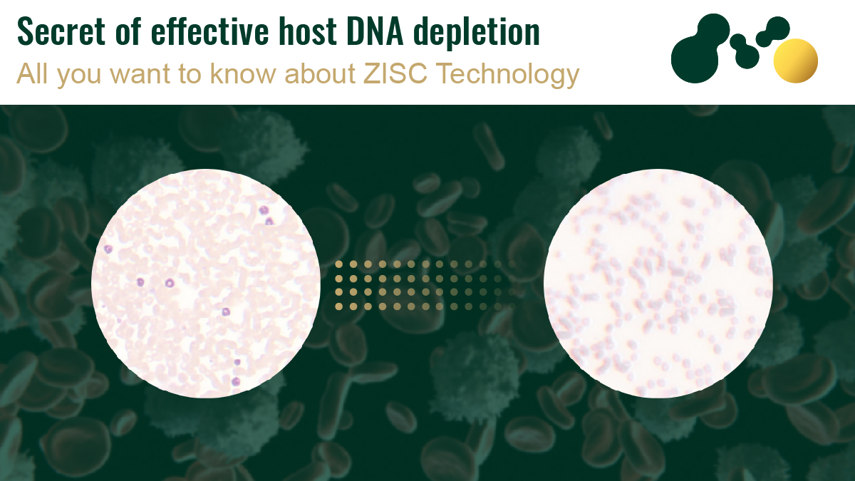
Devin™ filter, developed by Micronbrane Medical, uses a patented Zwitterionic Interface Ultra-Self-Assemble technology that can deplete 95% of human nucleated cells from biological samples in only 5 minutes. This depletion technology stands out among its competitors due to its capacity to deliver representative samples (with minimal interferences on microbial composition), with highly enriched fractions of bacteria and viruses and with minimal loss of key elements of the target microbiome (Fig. 1).
Figure 1. Comparison of contemporary host DNA depletion techniques with the Devin™ proprietary technology.
Devin™ filter is part of a pipeline of products developed by Micronbrane Medical that can expedite pathogen detection, using various next-generation sequencing platforms, with lower costs per sample and, ultimately, with multiple benefits to patients and healthcare providers alike. However, being an easy-to-use approach that requires non-specialized handling requirements, Devin™ filters also have the potential to act as a robust sample processing methodology to be coupled upstream of the available POC technologies for pathogen detection in sensitive contexts.
Advantages of the Devin™ filters for POC clinical diagnosis
Ready-to-use solution
Devin™ filters come 100% integrity tested, individually packaged and sterilized directly out of the box, requiring only a single syringe to operate with minimal handling requirements
ISO certified
Devin™filters are certified by ISO 13485 and are compliant with the best-practices of the medical industry
Highly versatile:
Devin™ filters can be used with whole blood, plasma and other body fluids (serum, swabs and washed) with low sample inputs (< 10 mL) and with quick processing times (up to 5 min)
Ensures reproducibility
Devin™ filters can ensure the traceability and reproducibility of the results by serving as a robust and effective sample preparation methodology for confirmatory genomic analyses (via NGS-based pathogen detection)
Devin™ filter is a fully validated and proprietary host depletion technology that can facilitate upstream sample prepping for POC clinical diagnosis. By being an out-of-the-box ready-to-use sample preparation solution, Devin™ ensures a quick and easy filter-based host depletion protocol that delivers high-quality biological samples compatible with most POC methodologies, while also ensuring full compliance with the best-practices in the medical industry.
In addition to this, Devin™ filters offer a unique opportunity to streamline the comparison of POC diagnosis results with the results obtained by more robust genomic approaches, namely NGS-based techniques, and can do so in combination with PaRTI-Seq™, another Micronbrane Medical product for NGS library prep, by enabling fast, effective and cost-efficient pathogen detection with reduced costs and very fast sample turnover. For more information on this, please check: OnePager
References
Pasquale, S., Gregorio, G. L., Caterina, A., Francesco, C., Beatrice, P. M., Vincenzo, P., & Caterina, P. M. (2021). COVID-19 in low-and middle-income countries (LMICs): A narrative review from prevention to vaccination strategy. Vaccines, 9(12), 1477. https://doi.org/10.3390/vaccines9121477
Roy, S., Arshad, F., Eissa, S., Safavieh, M., Alattas, S. G., Ahmed, M. U., & Zourob, M. (2022). Recent Developments towards Portable Point-of-Care Diagnostic Devices for Pathogen Detection. Sensors & Diagnostics. https://doi.org/10.1039/D1SD00017A
Wu, T. F., Chen, Y. C., Wang, W. C., Fang, Y. C., Fukuoka, S., Pride, D. T., & Pak, O. S. (2018). A rapid and low-cost pathogen detection platform by using a molecular agglutination assay. ACS central science, 4(11), 1485-1494. https://doi.org/10.1021/acscentsci.8b00447
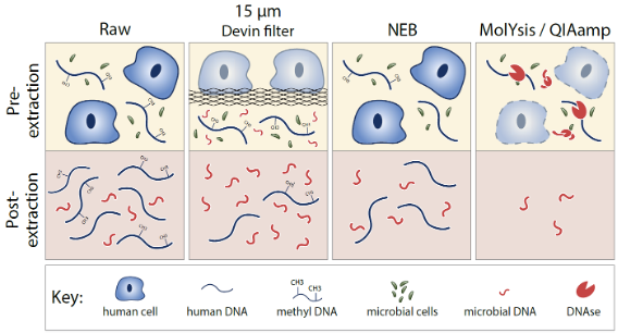
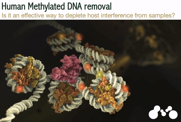
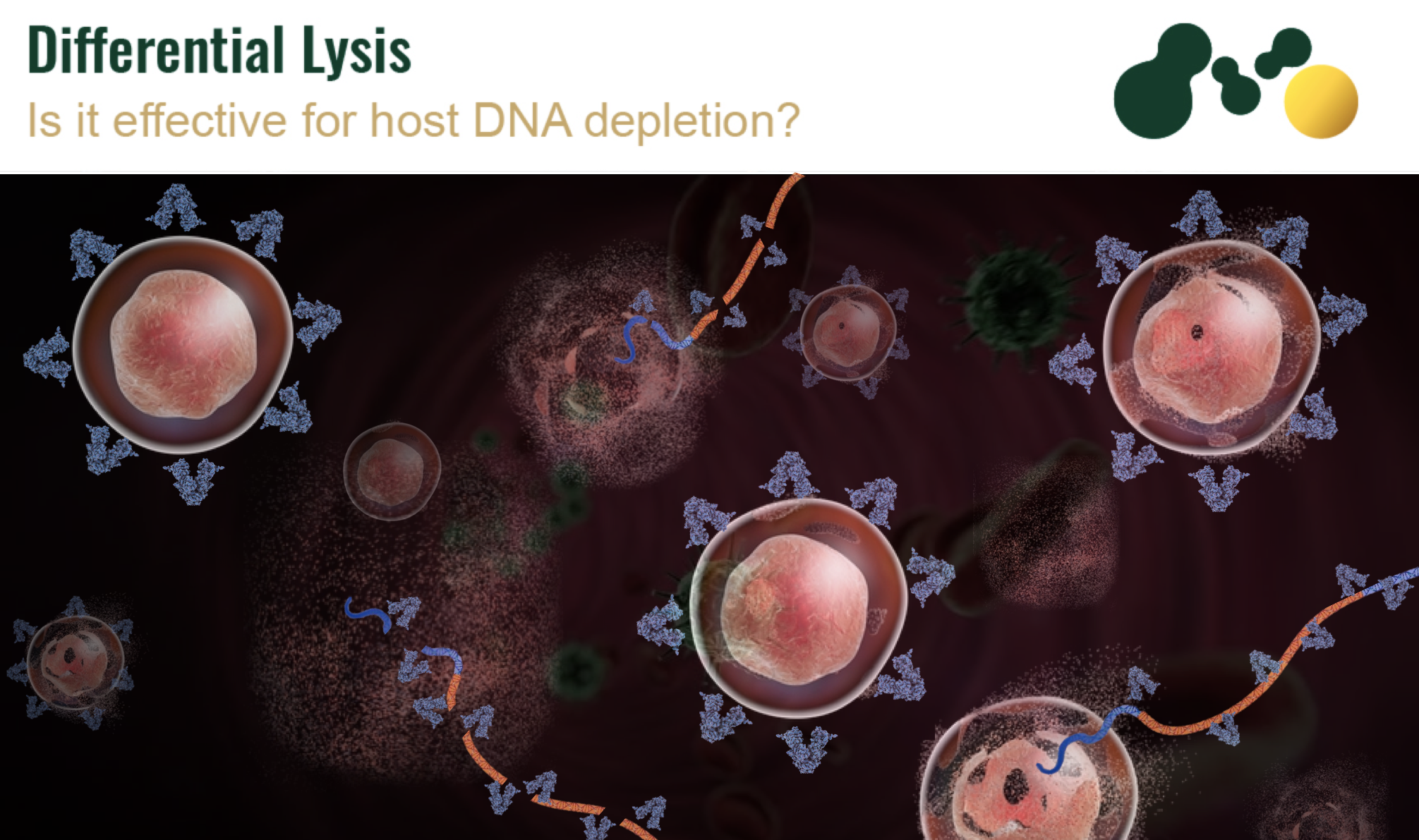
One of the constraints in sequencing-based methods of rapid pathogens detection is the presence of human DNA in the DNA samples. The size of human genome (3 billion bp) is very huge compared to the bacterial genome (average 4 million bp). So, the presence of a few human cells in the samples can result in overwhelming background noise in sequencing-based methods of microbial identification. So, there is a need to remove human DNA from the specimens. Without this the majority of the sequencing results would be generated from host instead of pathogen DNA. Nowadays there are several methods of human DNA removal, including differential cell size- based methods, chemical/enzymatic treatment methods etc. (Fig. 1) In this article we overview human methylated DNA removal methods and compare their efficacy with recently launched Devin® filter from Micronbrane Medical.
Figure 1. Contemporary Depletion Techniques
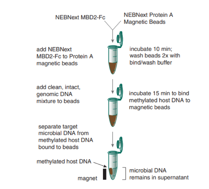
Abundance of methylated CpG domains in human DNA and extreme scarcity of CpG methylated sites in microbial DNA is utilized in many commercial methods to deplete host DNA via methylation. The commercial microbial DNA enrichment kits specifically target these sites to remove CpG human methylated DNA from clinical specimens. The method usually uses MBD2 (Methylated CpG specific binding protein) coupled with Fc region of human Immunoglobulin G. The Fc region of IgG binds with Protein A, that is coated on the magnetic beads. This is how human methylated DNA is selectively removed with the application of magnetic field leaving the non-methylated microbial DNA in the sample (Fig. 2).
Figure 2. A flow diagram showing removal of CpG methylated human DNA for microbial enrichment
New England Biolab’s NEBNext Microbiome DNA Enrichment kit utilizing method above claims to be able to remove human methylated DNA up to 94 % and increases the reads from 8 to 43 folds.
However, the efficacy of this method of microbial DNA enrichment has been tested in a research study which highlights certain limitations of this method (1). NEBNext® Microbiome DNA Enrichment kit was used for microbial enrichment from sino-nasal swabs. The study showed reproducible results (Confidence interval <100%), but the samples treated with NEBNext® Microbiome DNA Enrichment kit produced insufficient sequences for subsequent downstream analysis. Another drawback of this method reported is that the microbial community profile can be altered due to the magnetic field mediate removal of host DNA. The NEBNext Microbiome DNA Enrichment Kit can remove up to 95% of the human methylated DNA, but there are no significant increases in the amount of microbial DNA extracted as compared with control groups where microbial DNA was extracted using manual methods. Major limitation of this method is that it is expensive and requires high molecular weight genomic DNA input (> 15kb). This method is not suitable for specimens like saliva, serum, urine, plasma and samples that are likely to harbor no cellular portion (1).
The methylation dependent bacterial DNA enrichment requires pre-extraction of whole DNA from the clinical samples. The samples that do not have high cellular count such as saliva, urine and nasal swabs will result in small quantity of DNA. The minimum amount of DNA required for efficient removal of host DNA through CpG domains requires more than 3kb DNA, to which MBD-linked conjugated magnetic beads can bind. The low starting DNA materials result in inefficient binding of magnetic beads which in turn increase the background noise during sequencing and metagenomic analysis (5).
Other methods of microbial DNA enrichment include immunoprecipitation of host DNA that is mediated with methyl specific restriction enzymes. This method can remove human DNA up to 94% (2) but it requires long enzyme incubations hence no commercial kit working on this principle has been introduced into the market yet.
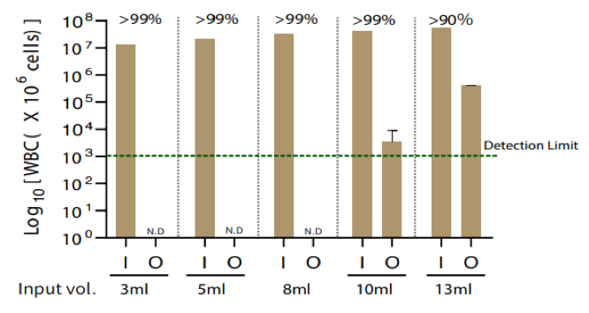
In comparison with human methylated DNA removal Devin® filter utilizes completely new technique of ZISC technology. Zwitterionic Interfaced Self-assemble Coating (ZISC) in Devin® membrane specifically binds with human leukocytes irrespective of the filter pore size and retain them without clogging the pores. The Devin® can deplete over 95% of human nucleated cells within just 5 minutes and possesses over 99% passing microbial efficiency (Fig. 3). Both bacterial cells and viral particles can easily pass through the filters (Fig. 4). Combined with PaRTI-Seq® it may increase microbial reads in the sample by 10-1000 folds compared with unfiltered samples. It is an efficient method for all types of samples, as it does not require pre-extraction of DNA so there is no minimum limit of DNA required for microbial DNA enrichment (3).
Figure 3. Leukocyte reduction efficacy.
Figure 4. Microbial passing efficacy of Devin® filters
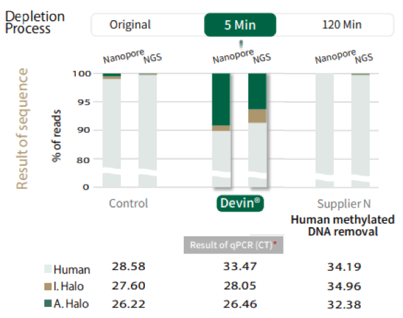
The NGS Nanopore sequencing and qPCR analysis of microbial DNA extracted via Devin® shows significant reduction in host contamination and improved microbial reads in NGS sequencing analysis as compared to methylation dependent human DNA removal and differential lysis method (4) (Fig. 5).
Figure 5. Comparison of efficacy of results of depletion method via NGS Nanopore sequencing and qPCR.
The comparison of microbial DNA extraction with Devin® and methylate dependent microbial DNA enrichment has been summarized in Table 1 below.
Table 1. comparison of Devin® and methylation dependent microbial enrichment method
Methyl dependent Microbial DNA enrichment
Microbial DNA enrichment with Devin®
This method takes 120 minutes to complete the enrichment process and prior DNA extraction is needed for further treatment with MBD2-Fc protein.
Can remove over 95% of human DNA contamination within just 5 minutes.
Microbial DNA profile can be altered due to application of magnetic field.
No alterations in microbial DNA profile, because sample is not treated with magnetic beads.
Does not produce sufficient sequences for downstream analysis.
Sufficient sequences are produced for any type of downstream analysis.
No suitable for samples i.e., saliva, serum urine and plasma.
Suitable for all types of specimens.
CONCLUSION
Devin® microbial DNA enrichment method is an advanced method to remove background noise created due to human DNA in sequencing, metagenomic analysis and PCR based microbial detection in clinical samples. This method has high efficacy (99%) and takes significantly less time (5 minutes) as compared to methylation method which requires more than 120 minutes to process sample for microbial enrichment. Furthermore, there are chances of DNA damage in methylation dependent method because Host DNA is removed under the influence of magnetic field, which can greatly alter the bacterial profile. Devin® microbial DNA enrichment method promises damage free extraction of bacterial DNA.
REFERENCES
Mackenzie, B. W., Waite, D. W., Biswas, K., Douglas, R. G., & Taylor, M. W. (2018). Assessment of microbial DNA enrichment techniques from sino-nasal swab samples for metagenomics. Rhinol. Online, 1, 160-193.
Glassing, A., Dowd, S. E., Galandiuk, S., Davis, B., Jorden, J. R., & Chiodini, R. J. (2015). Changes in 16s RNA gene microbial community profiling by concentration of prokaryotic DNA. Journal of microbiological methods, 119, 239-242.
Novel Human Cell Depletion Method Enables Rapid Pathogen Identication by Next Generation Sequencing.
Clarisse A. Marotz, Jon G. Sanders, Improving saliva shotgun metagenomics by chemical host DNA depletion; 2018 February 27
Feehery, G. R., Yigit, E., Oyola, S. O., Langhorst, B. W., Schmidt, V. T., Stewart, F. J., … & Pradhan, S. (2013). A method for selectively enriching microbial DNA from contaminating vertebrate host DNA. PloS one, 8(10), e76096.
Detection of pathogenic DNA from blood samples is essential in diagnosing bacterial, fungal, and viral diseases. Still, the amount of genetic information found in analyzed samples is extraordinary. Interference from host DNA decreases sensitivity for microbial detection and represents a significant drawback in identifying the pathogenic microorganism1.
What are the current lysis depletion methods, and how do they work?
Chemical or enzymatic techniques
These methods imply using a lysis solution such as a saponin and DNase treatment for the host DNA depletion. Chemical and enzymatic techniques decrease up to 99.9% or 105 folds of the human DNA in the samples to be analyzed. Saponin depletion can effectively remove the human genomes while preserving most pathogenic bacterial genomes. However, the pathogenic genome does not remain completely unaltered. This method requires 5–6 h from sample collection to pathogen classification.
Osmotic lysis
In this method, the lysis process is followed by propidium monoazide (PMA) or benzoate treatment to reduce host reads and increase microbial reads in the analyzed samples. Currently available lyse kits declare a host read decrease as low as 8 to even 5%. The required time just for lysis ranges from 40 to 100 minutes. One of the main advantages of osmotic lysis and PMA treatment methods is that this method requires fewer steps than other enzymatic methods and is also more cost-efficient2, 3.
Devin® filter
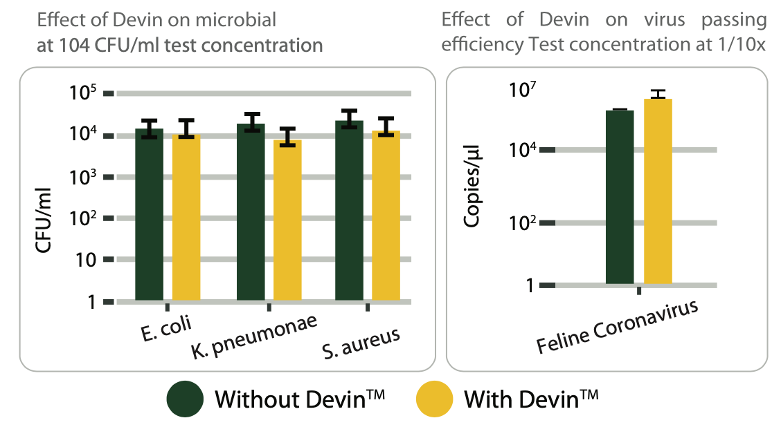
Devin® filter uses bio-compatible membranes to separate blood components and selectively remove up to 99.99% of white blood cells from the filtered sample. Devin filters do not interfere with the bacterial, viral, and fungal communities during the filtration process. As such, the pathogenic agents responsible for infections can be detected in the processed sample through nucleic acid isolation and amplification techniques. For testing, blood samples obtained from healthy donors were enriched with 10^4 Genome Copies/mL spike-in control (ZYMO Research) and then run through the Devin® filter. Results showed a quicker processing time and better enrichment results when compared to traditional methods4.
What are the limitations of currently used depletion methods, and what are the alternatives?
Bacterial cell wall structure variates from one species to another and significantly influences the lysis process. Consequently, the obtained results can be distorted by the bacterial cell lysis efficiency, and the relative abundance of bacteria in the analyzed sample can be misread. Studies developed on standardized microbial DNA samples revealed that the methods currently used to extract bacterial DNA can yield significantly different results.
The techniques observed to recover the lowest bacterial DNA used high temperature, ASL buffering solution, and protein kinase K for the lysis process. On the other hand, the highest microbial DNA yield was obtained on a method that used phenol-chloroform-isoamyl alcohol for extraction on which bacterial recuperation was 5.7, 5.4, and 3.3-fold higher on average for bacteria such as S. aureus, Pr. acnes, and C. tuberculostearicum. Saponin depletion methods can affect some microorganisms, such as Streptococcus pneumoniae, an important pathogen that can cause severe disease in humans56. Further, in the osmotic lysis, denaturation and disruption induced by PMA in the structure of proteins and nucleic acids can also affect extracellular bacterial DNA, lyse the bacteria and reduce the pathogenic community. Another critical limiting factor is the removal of intracellular viral material. Also, the high number of steps required to process a sample can affect the viability of the pathogenic community2, 3.
Devin® filter is among the best alternatives to currently used methods. The main advantage is that the technique does not require the usage of any chemical compound or lysing agent. Devin filters deplete blood samples of 99.99% of WBC and allow the unaltered passing of microorganisms. Compared to other tests, the depletion method of Devin filters is ideal for microbial enrichment of whole blood and different types of body fluids that require further metagenomic tests4.
Depletion techniques provide a new approach for rapidly identifying pathogens in clinical microbiology. Depletion methods have a higher sensitivity and require less time to establish a correct diagnosis than a clinical culture. Although highly reliable, depletion methods still have a series of critical shortcomings in depleting host genetic information, preserving pathogenic DNA, and ruling out a pathogenic microorganism as a causal agent for an infection. Techniques such used in Devin® filter overcome these limitations and could be key to cell depletion and clinical microbiology.
Reference
Heravi, F. S., Zakrzewski, M., Vickery, K., & Hu, H. (2020). Host DNA depletion efficiency of microbiome DNA enrichment methods in infected tissue samples. Journal of Microbiological Methods, 170, 105856. https://doi.org/10.1016/j.mimet.2020.105856
Charalampous, T., Richardson, H., Kay, G. L., Baldan, R., Jeanes, C., Rae, D., Grundy, S., Turner, D. J., Wain, J., Leggett, R. M., Livermore, D. M., & O’Grady, J. (2018). Rapid Diagnosis of Lower Respiratory Infection using Nanopore-based Clinical Metagenomics. Rapid Diagnosis of Lower Respiratory Infection Using Nanopore-Based Clinical Metagenomics. https://doi.org/10.1101/387548
Hasan, M. R., Rawat, A., Tang, P., Jithesh, P. V., Thomas, E., Tan, R., & Tilley, P. (2016). Depletion of Human DNA in Spiked Clinical Specimens for Improvement of Sensitivity of Pathogen Detection by Next-Generation Sequencing. Journal of Clinical Microbiology, 54(4), 919–927. https://journals.asm.org/doi/10.1128/JCM.03050-15
Wu, N., Ranjan, P., Tao, C., Liu, C., Yang, E., He, B., Erb-Downward, J. R., Bo, S., Zheng, J., Guo, C., Liu, B., Sun, L., Yan, W., Wang, M., Wang, W., Wen, J., Yang, P., Yang, L., Tian, Q. Shen, N. (2021). Rapid identification of pathogens associated with VAP by Nanopore sequencing. Respiratory Research, 22(1). https://doi.org/10.1186/s12931-021-01909-3
Yuan, S., Cohen, D. B., Ravel, J., Abdo, Z., & Forney, L. J. (2012). Evaluation of Methods for the Extraction and Purification of DNA from the Human Microbiome. PLoS ONE, 7(3), e33865. https://doi.org/10.1371/journal.pone.0033865
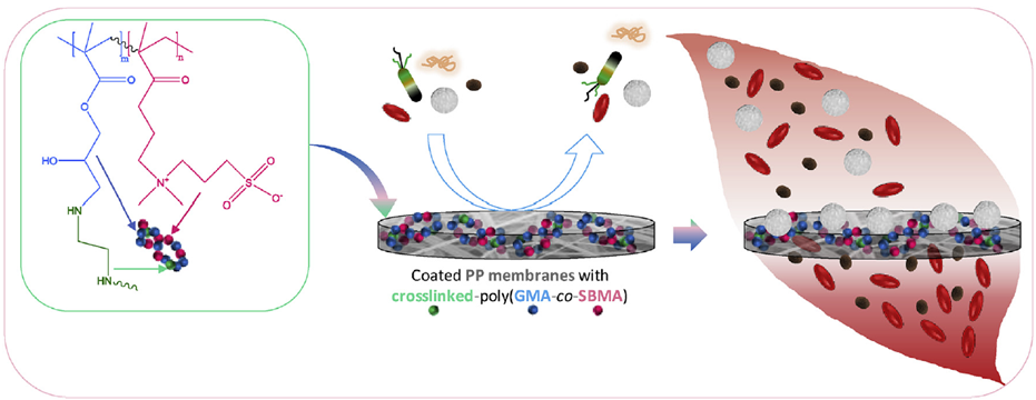
Blood-compatible membranes are among the most effective and cost-saving tools used to separate blood components. However, polypropylene (PP) membranes currently used in blood-contacting devices are prone to biofouling by host blood cells, proteins, and bacteria. Host contamination is one of the most critical bottleneck factors that limit the usage of PP membranes in clinical microbiology. The development of a filtering membrane resistant to biofouling is essential not only in clinical microbiology but also in blood filtration processes, water treatment, and the food industry.
Compared to other types of materials, zwitterionic networks have higher stability in complex mediums. More importantly, zwitterions provide strong hydration, and the modification process results in better hydration of the membranes. This characteristic acts as an energetic barrier against adsorption and biofouling by proteins, bacteria, and blood cells. Essential advantages of developing the zwitterionic membranes are that the method does not involve irradiation, oxygen plasma treatment, or UV irradiation.
Protein, bacteria, and blood cell biofouling prevention
Protein biofouling observed in commercial membranes promotes the adsorption of bacteria and blood cells. One of the most essential proteins found in blood samples is fibrinogen, a vital plasma protein involved in the formation of clots. Fibrinogen adhesion during filtration processes leads to white blood cell adhesion, deformation, and cellular death. The results obtained from Zwitterionized membranes indicated a drastic decrease in fibrinogen adsorption from 6.4 to 0.9 μg/cm2.
Zwitterionized membranes were tested for resistance to Escherichia coli, a bacterium commonly responsible for medical infections such as urinary tract infections. The bacterial adherence value was set to 100%, and results revealed a decreased E.coli adherence to the lowest relative measured attachment of 3 ± 1%.
How modified PP membranes remove white blood cells from blood samples
Compared to the commercial membranes where the pore size is reduced, the method through which the membranes are developed preserves the large pore size and porosity (Fig.1). These traits are essential for blood filtration processes, red blood cell preservation, and retention of white blood cells.
Fig. 1. The effect of coating poly(GMA-co-SBMA) with/without cross-linking agent (EDA) on (a) the structure, (b) pore size, and (c) porosity of modified PP fibrous membranes.
When a blood sample is filtrated through the Devin filters, white blood cells remain in the retentate or the part that does not pass through the membrane. Not only this, as compared to other types of membranes, the shape of white blood cells remains unaltered, and cells are still viable.
Multiple tests revealed that the modified PP membranes were highly efficient in selectively removing 99.99% of white blood cells from the filtered blood sample without affecting the erythrocyte concentration (Fig. 2).
Fig. 2 Schematic presentation of leukocyte removal from whole blood during blood filtration using modified PP membranes with a cross-linked poly(GMA-co-SBMA) polymer
The permeate, or the part that passes through the membrane, revealed a concentration of 0.07 103 cells/μL after filtration, while the initial concentration was 9.39 103 cells/μL. In comparison, unmodified membranes do not permit blood flow, and the commercially available hydrophilic membranes are not suitable barriers for white blood cells (Fig. 3).
Another advantage of poly(GMA-co-SBMA)-coated membrane is that the red blood cell concentration remains unchanged after filtration. This result indicates that these filters can be used to prepare RBCs-rich blood fractions, essential to patients suffering from intense bleeding after physical trauma.
Fig. 3 Comparison of poly(GMA-co-SBMA)-coated membranes with virgin unmodified PP and 3 or 5 layers of commercial hydrophilic membranes. (a) Whole blood filtration; (b) Concentration of red blood cells and white blood cells in the permeate (dotted lines) and white blood cells removal ratio determined from the white blood cells concentration in whole blood
Overall poly(GMA-co-SBMA)-coated zwitterionic membranes showed a significant improvement in blood compatibility with all analyzed samples. Multiple tests showed that compared to commercial membranes poly(GMA-co-SBMA)-coated zwitterionic membranes proved higher resistance to biofoulants adsorption and ideal cell preservation proprieties. The results and characteristics of the modified zwitterionic membranes indicate that this technology effectively reduces leucocytes in blood samples and removes host contamination from body fluids like whole blood. Micronbrane holds patent for this unique technology and utilizes it in its novel Devin® filter. PaRTI-Seq® built upon Devin® filter can provide precise test results within less than 24 hours upon sample receival.
To provide the best experiences, we use technologies like cookies to store and/or access device information. Consenting to these technologies will allow us to process data such as browsing behavior or unique IDs on this site. Not consenting or withdrawing consent, may adversely affect certain features and functions.
Functional Always active
The technical storage or access is strictly necessary for the legitimate purpose of enabling the use of a specific service explicitly requested by the subscriber or user, or for the sole purpose of carrying out the transmission of a communication over an electronic communications network.
Preferences
The technical storage or access is necessary for the legitimate purpose of storing preferences that are not requested by the subscriber or user.
Statistics
The technical storage or access that is used exclusively for statistical purposes.The technical storage or access that is used exclusively for anonymous statistical purposes. Without a subpoena, voluntary compliance on the part of your Internet Service Provider, or additional records from a third party, information stored or retrieved for this purpose alone cannot usually be used to identify you.
Marketing
The technical storage or access is required to create user profiles to send advertising, or to track the user on a website or across several websites for similar marketing purposes.